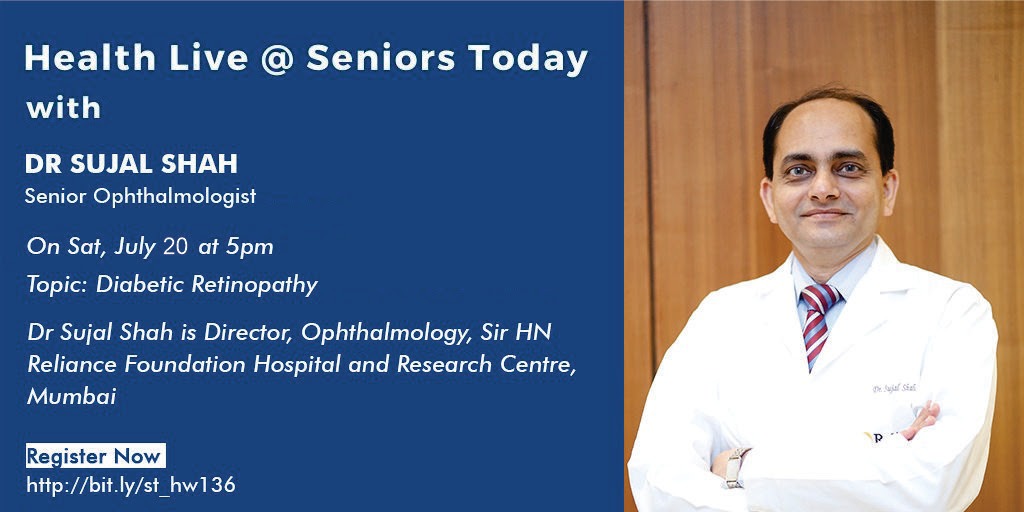
Dr Sujal Shah, Director, Ophthalmology, Sir HN Reliance Foundation Hospital and Research Centre.
After completing his MBBS, Dr Shah completed DO at Sankara Nethralaya, Chennai and also completed DNB (Ophthalmology). Then, Dr Shah pursued fellowship in Refractive Surgery Techniques at Doheny Eye Institute-USC, Los Angeles, Cleveland Clinic Foundation, and Jules Stein Eye Institute-UCLA, Los Angeles in the US.
Dr Shah has a rich experience of 20 years in the field of Ophthalmology. Apart from his association with our hospital, he is also a Consultant Ophthalmologist at Samyak Drishti-the Vision Institute, Mumbai. Earlier, he worked as a Consultant at Lilavati Hospital & Research Centre, Bhatia General Hospital & Research Centre, Conwest Jain Clinic, Sir H N Hospital & Research Centre, Surya Eye Tech SuperSpeciality Eye Hospital, Aditya Jyot SuperSpeciality Eye Hospital and Divyadrashti SuperSpeciality Eye Hospital. He started the Roshni Eye Bank and Tissue Processing Centre at Lilavati Hospital and was in charge of the Eye Bank at Conwest Jain Clinic and Bhatia General Hospital. He was on the advisory board of the Eye Bank Coordination & Research Centre, the Central Eye Bank and Processing Centre in Mumbai.
He specialises in cornea, cataract and refractive surgery. He has vast surgical experience in penetrating and lamellar corneal transplants, cataract surgery, refractive cataract surgery with premium lenses (MICS, Aspheric, Toric, Multifocal, Multifocal Toric, Trifocal & Trifocal Toric IOL’s) and the complete range of refractive surgeries (Basic LASIK, custom LASIK, Wavefront Guided LASIK, Topoguided LASIK, SBK, BladeLess Femto-LASIK and No blade No Flap ReLEx Smile, Phakic IOL’s, Refractive Lens Exchange, Bioptics, Corneal Collagen Crosslinking, Corneal Collagen Crosslinking with Surface Ablation and Blended Vision for reading glasses). Dr Shah was the first to start the commercial application of ReLEx Smile in India. He was the first to start Laser Blended Vision for Presbyopia (reading glasses in India). He has conducted clinical trials in India for advanced treatment for the MEL 80 excimer laser of Carl Zeiss, Germany.
He was on the editorial board of the Indian Journal of Ophthalmology. He is a reviewer for the Journal of the American Academy of Ophthalmology, American Journal of Ophthalmology, Journal of Refractive Surgery, Journal of Clinical Ophthalmology & Research. He conducts training courses for Ophthalmologists in LASIK, BladeLess LASIK and Phakic IOL’s. He mentors candidates for their doctoral thesis in Ophthalmology.
- Diabetes is a complex group of metabolic problems and it can be insulin or non- insulin dependent diabetes.
- Diabetes causes macrovascular (large vessel) or microvascular (small vessel) problems in different organs of the body. It can affect any part of the body including the eye.
- Very often, when proliferative changes start in the eye, the kidneys are also involved around the same time.
Prevalence:
- India has approximately 7 million diabetics. It is predicted to touch 125 in the next 20 years. Which means 1 in 5 adults is diabetic. And diabetic retinopathy is an important ocular impairment in diabetics.
- The prevalence of diabetic retinopathy is in 12.7% of diabetics and 4% of them have severe/ visually threatening diabetic retinopathy (VTDR).
Risk factors of VTDR include:
- Duration of disease
- Glycemic control (increased risk in patients with poor control)
- Concurrent disease such as hypertension, kidney problems, hyperlipidemia, obesity
- Smoking
- Pregnancy
- Loss of some of the cells in the periphery of the blood vessels that starts to make the cell wall weak leading to plasma leakage through that and that leads to retinal oedema. It can also lead to cholesterol deposition leading to formation of hard exudates.
- Sometimes, when there is no leakage but the capillary wall is weak, there can be bulking because of that leading to a micro aneurysm. And this can sometimes cause leakages or intra- retinal haemorrhages.
- There is also basement membrane thickening leading to endothelial damage.
- Deformed RBCs, platelet aggregation can also lead to microvascular occlusion/ blockage in thermal blood vessels. Therefore, there is hypoxia (less oxygen supply through the blood to the parts of the retina). Hypoxic retina secretes Vascular Endothelial Growth Factor (VEGF) which stimulates the growth of new blood vessels.
Based on the severity, Diabetic Retinopathy can be of 2 types:
- Proliferative: new blood vessels are forming
- Non proliferative: no new blood vessels are formed. This can further be classified into:
- Mild: normal macula and optic disc with areas of leakages called cotton wool spots, some haemorrhages, outpouching of blood vessels called micro aneurysms.
- Moderate: some sacculations in the blood vessels are seen, bleeding of blood vessels, ischaemia, larger yellow (cotton wool spots) spots are seen
- Severe: the same aforementioned changes are seen but more severe in presentation. More cotton woo, spots and exudates are seen. Intra retinal haemorrhages are also seen.
- Very severe
- And this can be associated with clinically significant macular oedema
- Till the centre of the retina (macula) is not involved the image is formed and there is no clinical complaint. Which means, until a regular examination is not performed, more often than not, something like this often goes undetected.
- When you have clinically significant macular edema (CSME), the above mentioned changes in the centre part (macula) gets thickened leading to deterioration in the quality and quantity of vision.
- The next stage is when new blood vessel formation starts to occur. The newly formed blood vessels are very thin and fragile and a form of mild shock/ trauma can cause these vessels to rupture leading to bleeding/ haemorrhage in the eye which can affect the vision.
- When new blood vessels are formed, there can also be fibrous proliferation and as fibrous proliferation tracts the retina, it can cause a part of the retina to get lifted which is called retinal detachment.
Causes for visual loss:
- Non clear vitreous haemorrhage
- Neovasuclar glaucoma
- Retinal detachment
- Macula ischemia
- Clinically significant macular oedema
Preventive measures to avoid/ prevent these changes:
- Till you do not get a haemorrhage/ macular oedema does not set in, the vision is maintained. Which means that early detection needs to be done.
- Routine screening of the retina after dilation of the eye.
- Thus, routine evaluation and examination helps in early detection and faster treatment and intervention. With this, you can avoid the progression of the disease to visual impairment/ loss. Which means you can be diabetic for life but your vision will be maintained.
- Good glycemic control
- Control over other comorbid conditions
Severe and very severe DR requires close monitoring by the ophthalmologist.
CSME and proliferative DR require close follow up.
In severe and very severe non proliferative DR, we can do laser therapy to prevent loss of vision due to haemorrhage
In the early stages of proliferative DR, we can use lasers to prevent vision loss. This reduces the oxygen load on the retina and reduces the load on the retina.
For all diabetics, a yearly screening by fundoscopy is minimum and required.
The above mentioned ocular changes generally start 5 years after the onset of diabetes.


