Reading Time: 4 minutes
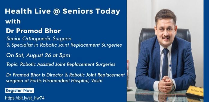
On 26 Aug, 2023, Seniors Today hosted its weekly Health Live Webinar. This week we had with us, a Senior Orthopaedic Surgeon and Specialist in Robotic Joint Replacement Surgeries, Dr Pramod Bhor who spoke on and answered questions about Robotic Assisted Joint Replacement Surgeries.
Dr. Pramod Bhor is Director – Orthopedics and Joint Replacement Surgeon at Fortis Hiranandani Hospital, Vashi.
He is a highly qualified doctor holding degrees in MBBS from BVPMC, Pune, and MS – Orthopaedics from Dr. D.Y. Patil Medical College and Hospital, Pune in 2006. Furthermore, he has received specialised training from several eminent national and international medical centres.
Academically, he has been teaching MBBS and PG students for the past 15 years and is a Professor of orthopaedics. He has many publications to his name in many national and international journals. He is the first surgeon in Navi Mumbai to perform Robotic knee replacement surgery.
Some of the services performed by Dr Pramod Bhor at Fortis Hiranandani Hospital are ACL Reconstruction, Arthroscopy, Hip Replacement and Resurfacing, Spinal Surgeries, Robotic Knee Replacement and Knee Replacement. He’s a member of the Bombay Orthopaedic Society, Navi Mumbai Orthopaedic Association and Maharashtra Medical Council, and many other orthopedic associations. He has done fellowships in Advanced Trauma and visiting fellowships in joint replacement in Singapore and Germany.
Joint replacement is a surgery which is usually done for severe osteoarthritis of the knee.
Every patient over the age of 55-60 years is going to have osteoarthritis. But not everybody needs surgery.
Surgery is necessary for:
- Patients who are in significant pain
- Patients who find no relieve in pain despite regular analgesic medicines
- Patients with no relief in pain/ other symptoms after physiotherapy
- Younger patients with secondary causes such as post traumatic, rheumatoid arthritis
Basic investigation and the investigation of choice which helps us diagnose osteoarthritis is an X ray of the joint. It shows us- joint loss, joint contour, joint space, sub chondral sclerosis and new bone formation.
Dr Bhor prefers an MRI for only the younger group of patients, who might have early osteoarthritis, medical joint arthritis- in this case the MRI is done to confirm that only the medial joint is involved and if the later joint is also affected and the extent of involvement, if any. This is done to see if a partial knee replacement will be a feasible option or not.
Management modalities for osteoarthritis of the knee:
- Medicines
- Intra articular injections
- Pain management techniques
- Taping
- Increase the muscular strength
- Surgical- arthroscopy, tibial osteotomies- not preferred these day, total knee arthroplasty- the most commonly done, when all other treatment modalities fail.
What is done in a total knee replacement surgery?
The damaged cartilage is shaved off, and a metal cap is placed over the femur, tibia and the space in between. While doing this, we balance the ligaments that are on the outer side and the inner side and correct the deformity.
Over the years there have been upgrades in the implants, implant designs, impact materials, methods and techniques of performing the surgery and the most recent upgrade is the robotics surgery.
The impact designs have also improved leading to better flexion and mobility for the patient. They can even squat as per Indian standards, however Dr Bhor does not advise squatting even then, because squatting, sitting cross legged puts more strain on the joint and decreases the life of the implant which is not ideal and thus, not advised.
Cruciate retaining implants are also available which helps in keeping the ligament intact because of which there is a more natural strength and feel to it.
Nowadays, we have the availability of hypoallergenic materials which also have a longer life, better stability and low friction.
A small scar does not necessarily mean minimally invasive. Minimally invasive surgery means-
- Reduced blood loss
- Lesser soft tissue damage/ dissection
This leads to faster rehabilitation and lesser postoperative complications and side effects.
The size of the skin incision does not matter, what matters is the soft tissue dissection and the size of the muscle dissected.
Pain management techniques in surgery are better in today’s times. Earlier, post operatively, the patients would be advised to stay in bed for about a weeks’ time and then start mobilising the knee.
Nowadays, the patients are advised to start moving the joint, flexing and extending the knee on the first day itself. This way the patient has better mobility, more confidence and lesser post operative pain and complications when mobility is started earlier.
Robotic knee replacement is the latest advancement we have seen.
The robot does the bone cutting, but the site and precision is decided by the surgeon.
This can be image based- after doing a CT, or it can be imageless, no CT scan is done prior to the surgery.
By doing the CT scan we can understand the bone anatomy better, we have a 3D picture. The CT scan can be viewed and a plan can be made one day prior to the surgery. This gives the surgeon a chance to do a virtual surgery one day prior to the actual surgery. Because of this, the surgeon can give better and consistent results.
Cuvis is a fully automatic robot.
Stryker is a semi automatic robot.
We perform robotic surgery to reduce human error while operating.
The robot has a robotic arm, which has a cutting device
And a console which tells us where the robot is going to cut, how deep and if it is going it correctly or not.
Optical tracker- these are sensors which we apply on the bone- femur and tibia. When these sensors are applied, the optical tracker detects where the femur and tibia are. Similarly there are trackers on the robot and robotic arm. This way, it knows where the robot is standing, where the robotic arm is cutting and how deep.
Planner is a software on the laptop where one day prior the plan for the surgery is made and virtually visualised.
While doing the surgery, the plan is fed to the computer.
The surgeon opens the joint, the robot has no role to play thus far. The points are marked on the femur and the tibial which is then read by the optical marker. This forms a 3D image.
This 3D image and the image of the CT scan are superimposed at the time of the surgery and this gives the surgeon a better and clearer picture on whether he wants to change his plan, the depth of his incisions, the site of his implant, etc.
Advantages:
- Better surgical planning
- Precision
- Better optical alignment
- Flexibility to change the cuts while operating depending on the patient
- Real time system monitoring


