If detected and acted upon on fast enough, a stroke patient can get back to a normal life, writes Dr Anirudh Kohli
Eighty-five-year-old Dr. Susil Chandra Munshi is one of India’s best-known cardiologists. He is a practicing interventional cardiologist at Mumbai’s Jaslok and Breach Candy Hospitals. Dr. Munshi has been President of the Indian Cardiology Society and was awarded the Padma Shri by the Government of India for his contribution to the field of medicine, specifically cardiology.
Ten years ago, he started to slur one evening. His speech became garbled and he just couldn’t speak. He also lost consciousness momentarily. His daughter rushed him to the Breach Candy Hospital. On arriving at the emergency medical services department, the doctors found that his right arm and leg had zero power. He had had a full-blown stroke. A CT scan was done immediately and that ruled out a hemorrhage, indicating an ischemic stroke. In view of this, he was given a clot-busting drug intravenously r-tPA. Within a short time, power returned to his arm and leg as well as his speech got better. “There was some brain swelling because of the stroke and I was a bit drowsy for two days but by Day 3, I had recovered fully,” he recalls. Upon investigation of how he got a stroke, it was discovered that he had a hole in his heart! “An embolus had crept up into my brain arteries, causing a critical blockage. This hole in the heart was from birth, but played up then. I got operated on by Dr Sudhansu Bhattacharya to close the hole in the heart and am absolutely fine now.”
What is a Stroke?
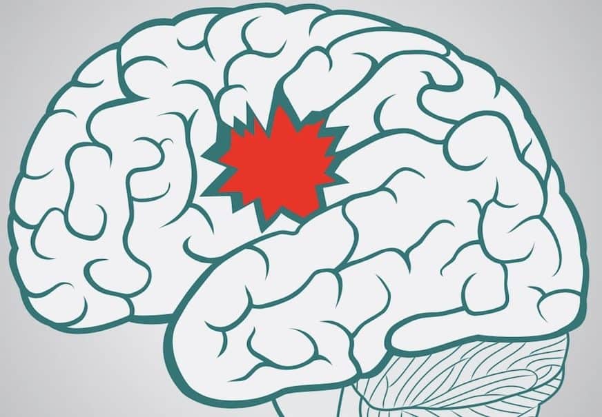
A stroke occurs when blood supply to a part of your brain gets reduced or interrupted thus depriving the brain of oxygen and essential nutrients. Within minutes, brain cells begin to die. That’s why a stroke is an emergency. The good news is strokes can be treated and prevented. Prompt treatment is crucial as early action can minimise damage to the brain tissue as well as complications.
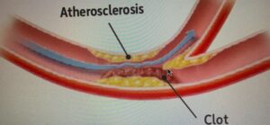
In a stroke, the arteries carrying blood to the brain are affected. There are two different types of strokes, ischemic and hemorrhagic strokes. Ischemic stroke is the most common, accounting for 80 per cent of strokes. In an ischemic stroke, arteries carrying blood to the brain get blocked. The brain tissue supplied by this blocked artery gets damaged. There are two main causes for ischemic stroke – a thrombotic or embolic stroke. In a thrombotic stroke, there is deposition of fatty material in the arteries supplying the brain especially in individuals with a high blood cholesterol level]. This fatty material plaque narrows the blood vessel and finally obstructs it blocking blood from reaching the brain (Figure 1). This most likely happens in the neck at a site known as carotid bifurcation or within the brain. An embolic stroke occurs usually as a result of a blood clot in the heart which is propelled upwards into the blood vessels supplying the brain to cause obstruction of the artery. It may also occur if a piece of fatty plaque in the neck arteries plaque breaks off and extends forward to block the arteries in the brain.
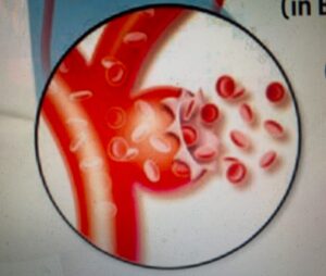
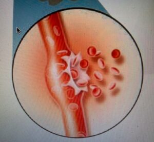
The second type of stroke is a haemorrhagic stroke, where blood vessels in the brain may burst. This occurs when a blood vessel in the brain bursts and spills blood into the brain tissue. There are many causes which make a blood vessel wall weak to burst. The most common reason is due to high blood pressure. Other causes are a weak spot in the wall of a brain artery known as an aneurysm. The wall bulges out into a thin bubble, as it gets bigger the wall may weaken and bursts. Another cause is rupture of a small tangle of thin walled abnormal vessels known as an arteriovenous malformation or over treatment/side effect of blood thinners. There are two types of hemorrhagic stroke – intracerebral haemorrhage when a blood vessel in the brain bursts and spills into surrounding brain tissue-damaging brain cells and the other type is subarachnoid hemorrhage where an artery near-surface of brain bursts and spills into space between surface of brain and skull.
Effects of a Stroke
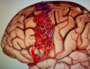
The human brain has different areas that control how the body moves and feels. When a stroke damages a certain part of the brain, that part may not work as well as it did before, cause problems with walking, speaking, seeing or feeling, pose challenges with basic self-care, bathing, dressing, eating, memory, emotions, and depression.
Diagnosis of a Stroke
How do you determine if you or someone is having a stroke?
Well, they may develop one of the more of the following signs or symptoms:
- Paralysis or numbness of face arm leg may develop suddenly. Often this happens on one side of the body – to test this, ask them to try to raise both arms and check if one side starts to fall. Also, ask them to smile, if one side of mouth droops. These are important signs of a stroke.
- Trouble seeing in one or both eyes – suddenly blurred or blackened vision in one or both eyes, or you see everything double
- The trouble with walking – may stumble, loss of balance, coordination, dizziness, altered consciousness
- A sudden severe headache may be accompanied with vomiting this is highly suggestive of a hemorrhagic stroke
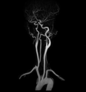
A quick mnemonic is FAST
Face Ask person to smile, if mouth one side drops
Arms Ask person to raise both arms – does one arm drift down or unable to raise one arm
Speech Slurred
Time Early diagnosis and treatment can reverse or minimise the effects of a stroke – a concept to remember is Time is Brain, so don’t waste time! Get to a hospital, go as quickly as possible.
At the hospital, the patient will be examined to confirm signs of stroke, history of past illness checked, blood pressure and other vitals checked and blood taken for examination.
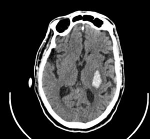
Stroke can hit anyone anywhere. Like it happened with Mr and Mrs Mehta (name changed to protect privacy). Married for some decades, this handsome elderly couple in their late 50s decided to trot the globe. On the itinerary was a cruise from Dubai to Mumbai. While watching television in the evening, Mr Mehta experience a sudden dizziness, slurred speech, swayed to the right and almost fell from his chaise longue. After primary care, the emergency code was activated and the captain of the cruise was alerted to dock the ship as fast as possible to Mumbai for further care.
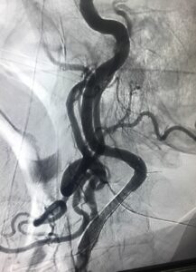
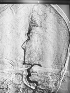
He reached the hospital after four hours of the stroke onset. I undertook an urgent MRI and MR Angiography Brain at the Breach Candy Hospital. One could see an occlusion of the right middle cerebral artery.
Since 2013-14, the physical removal of the thrombus (clot in blood tubes of the brain) from blood vessels of the brain have revolutionized the treatment of strokes.
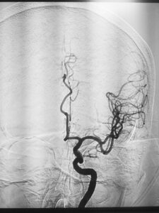
Mr. Mehta came in during the Golden Hours of a stroke (within eight hours of stroke onset). After the thrombus removal by Dr. Nishant Aditya, Neuro Interventionist at the Breach Candy Hospital, robust blood flow to the brain is restored.
Within 24 hours he recovered completely. His speech left side power and the balance returned to normal. On the sixth day, he went back home and the Mehtas resumed their itinerary.
Says Mr Mehta: “It was a life-changing experience. With urgent and diligent care by the team of doctors at Breach Candy, I was able to resume my social and business life in 10 days. Without this effort, I would have lost my active life. Thank you again, doctors!”
Dramatic Changes in Diagnosis
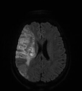
Recent advances in diagnosis and therapeutics of stroke over the last decade have dramatically changed the outcome and survival of stroke patients. To determine the appropriate treatment, the type of stroke has to be determined as well as the regions of the brain affected and the extent of damage. The best initial modality is a CT Scan.
This a computerised X-ray machine which will quickly scan your brain under a minute. All the patient has to do is lie down on the table; there is no dye injection required. The CT scan will tell if there is hemorrhage due to bursting of a blood vessel. If there is no haemorrhage, then an MRI is required to be performed immediately as it is very sensitive in demonstration of brain damage due to reduced blood supply.
The MRI will in the first minute itself tell conclusively if there is a stroke and the extent of damage. Once this is determined, the blood vessels are imaged, a process known as MR angiography. This maps the blood vessels from the heart to the minute vessels in the brain. This also does not require any dye, and takes about ten minutes. Care should be taken that the patient does not have a pacemaker as this is a contraindication for MRI. The MRI will demonstrate the extent of brain damage, as well as also if a major vessel is blocked. These pieces of information are very important in determining the treatment. If there is no major blockage of the vessels and no haemorrhage or contraindications, a clot-busting drug known as Alteplase r-tPA is given intravenously. This works by dissolving the clot and improving blood flow.
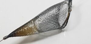
It must be administered within three hours of onset of stroke, for certain regions up to 4.5 hours. If there is block in a major vessel then the clot-busting drug may be used, but as the clot is usually large, the clot-busting drug may not dissolve the clot totally; in these cases mechanical thrombectomy is indicated. A catheter is passed from the groin to the block in the major artery, a small cage on a wire is passed through the catheter and the clot is sucked out into the basket. This should be done within six hours of onset of stroke, some may benefit even within 24 hours of onset of stroke.
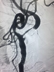
If there is a haemorhagic stroke, the key is controlling blood pressure, if it is due to blood thinners, medications may be given to try and reduce the blood-thinning, if due to an aneurysm then an endovascular procedure such as coiling or surgical clipping may be done. If AVM, then endovascular embolisation may be done or surgical excision
Life and Hope After Stroke
However, there is life and hope after a stroke. Though stroke will change your life in an instant, the quality of rehabilitation can help you recover close to your full potential.
Rehabilitation
This is very important as it can help you build your strength, capability and confidence, and help you continue your daily activities despite the effects of stroke. You may need to relearn or change how you live day to day for the most basic things in life such as dressing, eating, speaking etc. The goal of rehabilitation is to become as independent as possible. But rehabilitation comes from a combined effort by you, your loved ones and health care professionals. Both play an important role.
Healthcare professionals involved with rehab are essentially therapist and counsellors. Caregivers are very important as patient and caregiver need to be care partners. Patience and emotional support are very important components for caregivers. Stroke survivors feel tired as they have to work harder to make up for the loss of normal functions, and they usually have lack of energy and lack of motivation.
Rehabilitation should focus on
- Activities of daily living such as eating, bathing, toileting, grooming, and dressing – occupational therapy.
- Mobility, getting from bed to chair, walking, climbing stairs or using a wheelchair – physical therapy.
- Communication skills in speech and language – speech therapist.
- Cognitive skills such as memory or problem-solving.
- Social skills such as interacting with other people.
- Recreation skills, skills enjoyed before the stroke, as well as introducing new ones.
- Vocational rehabilitation evaluates your work-related abilities and helps you make the most of your skills when you return to work.
- Psychological functioning to improve coping skills and treatment and to overcome depression.
Rehabilitation is part-recovery, part-learning, essentially to adapt for deficits that may not fully recover. Adaptive strategies are the new normal!
Steps to prevent a stroke
It is very important to prevent a stroke from happening as not only the patient incapacitated but also the family and caregivers as a lot of support is required.
The key points in preventing a stroke are:
Blood pressure – Monitor your blood pressure; this is a silent killer that quietly damages blood vessels and leads to serious health problems.
Cholesterol – Control your cholesterol; the ideal level is 150 mg/dl.
Weight Loss – Lose weight if you need to. Get active and exercise daily – brisk walk and swimming are the simplest and best.
Smoking – If you smoke, you should stop, as smoking doubles the risk of stroke.
Drinking – if you drink alcohol, do so in moderation. Heavy drinking can increase your risk of stroke.
Diabetes – Diabetes increases the risk of stroke. Look at your diet, and increase vegetable and fruits component, as this will help manage diabetes.
Salt – Cut down on sodium intake.
Atrial fibrillation – Find out if you have atrial fibrillation, a condition where the heartbeat is irregular, that can lead to blood clots and cause embolic stroke. A simple examination of your pulse and ECG will detect atrial fibrillation.
Transient ischemic attack
This is also called a mini stroke. When an artery is blocked for a short time, blood flow initially slows down or stops and then the artery gets unblocked quickly or a new path opens up, blood flow is restored, symptoms last for a short time and then disappear, this is actually a warning that a full-blown stroke may occur in the near future. If you have had a transient ischemic attack please get investigated with an echocardiogram to see if there are clots in the heart, Holter monitoring to detect an abnormal rhythm, neck doppler to check for any plaque in the carotid arteries, MR angiogram to detect plaques in brain vessels. All these are treatable. If there are clots in the heart they can be dissolved with blood thinners, if there is an abnormal rhythm medication or a pacemaker can control it. If there are plaques in the neck vessels these can be treated with stenting or surgery. All these will prevent a stroke from happening. (A Holter monitor is a medical device that measures the heart’s rhythm.)
A good motto to maintain:
I will act FAST
I will make changes in my daily life that will help prevent a stroke
I will help my family and friends make changes that will prevent strokes
If detected on time and acted upon without delay, stroke patients can get back to their normal day-to-day lives soon enough. Reminisces Dr Susil Munshi: “I am back to opening up the blocked heart vessels of patients, and am thankful that my own blocked brain vessel was opened up. This was thanks to timely rushing to the hospital, discovering the stroke and receiving a clot busting drug. If all this was not diagnosed and treated, I would have been left with paralysis on the right side, I would not be able to do angiography and the complex angioplasties I do till today.”




Its find so comfortable and well explained to easy understand .
On 3rd Dec 2018, I had a massive ischemic stroke paralysing my full left side of body from head to toe. I was rushed to Hiranandani Hospital powai & was given the clot buster injection within an hour of stroke. I am lucky to be almost fully healed & am doing my normal activities. I always forward educational messages on stroke to help as many people as I can. Thank you
If possible try to send details in hindi
A very useful article.Thank you very much.
Quite educational article of high importance
Thanks Dr for this useful article
My wiife has suffered a stroke. She was treated at Rangadorai hospital, Bangalore. Though sha conscious she is always bed ridden. How to cure her completely?
Very knowledgeable description about strokes and remedial actions very well narrated thanks my wife did have left aneurysm on 29/12/18 she was in icy till Feb end discharged on24/3/19 is in semi state paralytic no speech techso my and pet feeding removed please pray for recovery
Thanks to you it is very help full to us in our future life without disturb.tks
Comments are closed.